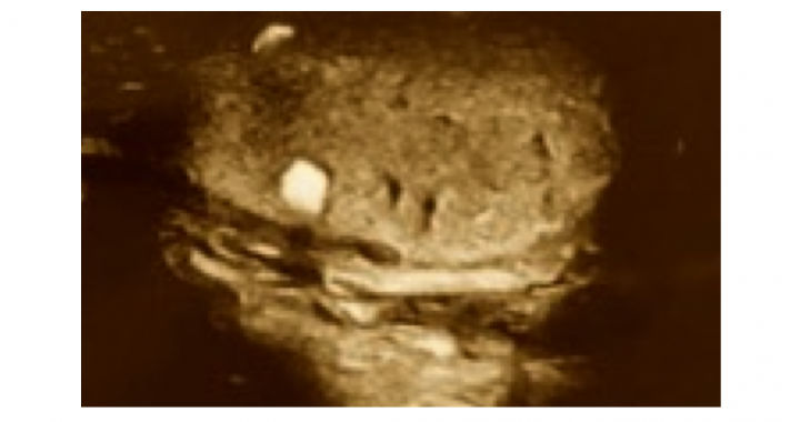
Testicular ultrasound is a diagnostic technique that allows to quickly and easily assess numerous testicular parameters that may be relevant for fertility treatment, but also for the general health of the man.
Ultrasound can assess the consistency of the testicular tissue, the presence of nodules or cysts, the alteration of vascular flow with the presence of varicose veins (varicocele), the accumulation of peritesticular fluid (hydrocele), the presence of hernias or the obstruction of the sperm passages that can cause azoospermia.
Testicular ultrasound is especially indicated in the following cases:
– Appearance of swellings or tumors at the testicular or scrotal level.
– Acute or chronic testicular pain
– Increase or decrease in the size of one of the testicles.
– Important alterations of the seminogram
– Erection problems
– Patients with previous testicular pathology in follow-up (calcifications, after removal of cysts or tumors …)
When we talk about alterations in the seminogram, the most frequent finding is the presence of a varicocele, which consists of the dilation of the testicular cord veins and which can explain up to 40% of cases of male infertility.
The decision to perform a surgical intervention for this varicocele will depend on its degree, as well as on the sperm alteration observed and the degree of improvement expected after the intervention. All of this, together with the recovery time for spermatogenesis after surgery, must be agreed with the patients before performing an intervention that may not improve their reproductive prognosis as much as we expect.
Other less frequent findings that are important to detect to favor a correct diagnosis and treatment are epididymal cysts and tumors, hydrocele, scrotal hernias or testicular cancer.

