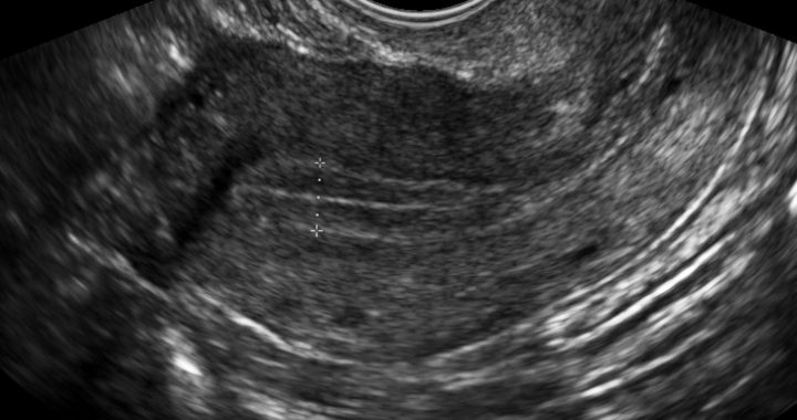
The vaginal ultrasound (VUS) is a painless imaging technique that allows evaluating the shape, size and activity of the ovaries, as well as the uterus and, under certain circumstances, the Fallopian tubes.
It is an indirect evaluation, as images are provided by ultrasounds and it is a fundamental tool for the diagnosis of infertility and in the monitoring of assisted reproduction treatments.
When performed at the beginning of the menstrual cycle, it allows indirect assessment of the number of antral follicles (follicles between 2 and 9 mm) in the ovary, which reflects the ovarian reserve.
During the monitoring of the natural cycle or during ovarian stimulation, it will allow us to measure and control the development of the follicles to determine the moment when they reach maturity.
Regarding the evaluation of the uterus, ultrasound allows evaluating its size and shape to determine the presence of fibroids, polyps, adenomyosis, which may be the origin of infertility and may require an specific treatment. For uterine evaluation, 3D ultrasound and hysterosonography (introduction of saline into the uterus at the same time as the ultrasound) can be combined and provide valuable information when it comes to uterine malformations with a negative impact on pregnancy .
For the evaluation tubal patency, ultrasound scan can identify the presence liquid accumulation, called hydrosalpinx, which interfere with their normal functioning. However, the best technique to visualize the tubes under US is the HYFOSY (Hysterosalpingo-Foam-Sonography) which uses a liquid foam to detect stenosis and blockages of the tubes. We will develop this technique later on.