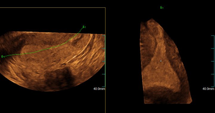
3- dimensional ultrasound has been widely used in obstetrics for the evaluation of fetal pathology but also for the visualization of the facial characteristics of the fetus before birth.
However, its application in gynecology has also been increasing over the last few years, as it is a very useful tool for evaluating certain uterine malformations trough the acquisition of a third plane that allows visualizing the structures in depth and with great precision.
This technique allows a more precise diagnosis of many pathologies, allowing a specific and adequate treatment for each patient and avoiding unnecessary delays and more invasive tests.
Two-dimensional ultrasound scans are usually performed in the gynecological consultation, but in the case in which there are doubts about the integrity of the female genital tract, a more detailed examination with 3D ultrasound can be requested.
3D ultrasound is performed in a similar way to conventional transvaginal ultrasound, although it may require a little more time for its correct evaluation and adequate image capture.
The advantage of 3D ultrasound is that the volumes obtained can be saved for later evaluation and even for discussion with other specialists in the most complex cases.

